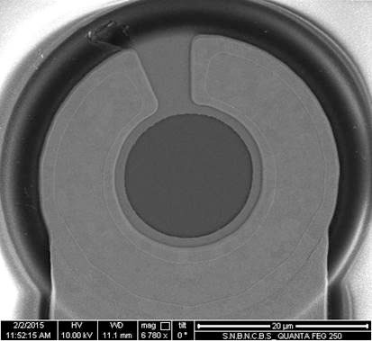
SEM/FESEM
SEM or Scanning Electron Microscope is an electron microscope which creates the image of a sample by scanning a focused electron beam on to it. The electron beam is generally scanned in a raster pattern and the detected signal combined with the beam position forms a high resolution image. In most common mode the SEM detects the secondary electrons emitted by the atoms of the sample excited by the focused electron beam. The FESEM (Field Emission SEM) uses a field emission cathode in the electron gun which produces a much narrower beam of electron compared to the conventional SEM for a wide range of electron energy. This results in a high spatial resolution as well as less charging effect. In FESEM the elemental analysis of a very small area of interest using Energy Dispersive X-Ray (EDX). It's possible to get high quality image at a low electron voltage (as low as 0.5kV) which produces minimum charging of the sample. So a conductive coating is often not required in the FESEM. As a low electron beam energy can be used, the surface damage is also minimum. Compared to conventional SEM, the FESEM produces a much clearer image.
